
Tuesday, September 30, 2008
To blog or not to blog...
Every so often I look at the hit counts for Talking Brains to see if it is worth continuing to post. If no one is looking, there's no point in posting. With over 3500 hits for the last month -- and probably at least some of these were on purpose -- I suppose it is worth continuing...

Shmoozing in Durham (UK)
If you happen to be in Durham this weekend, come and hang out at this party:
From Neuron to Language
States of the art in the cognitive neuroscience of language
A couple of departments at Durham are hosting a little symposium on this stuff, with the hope of stimulating philosophical discussion. Speakers include Richard Wise, Angela Friederici, Gary Marcus, Lolly Tyler, and some others.
Here is the idea: "All invited speakers are asked to take a step back from their day-to-day research and assess what has and has not been achieved in terms of an understanding of the brain basis of the human linguistic mind. The overall aim of the symposium is largely philosophical, insofar as we wish to understand the mind in terms of the brain, yet the approach is bottom-up in that we approach the issue from the experimentalist’s point of view."
If all of this happens with appropriate levels of lubrication (is gin a lubricant?), it could be quite entertaining. As you can predict, I will argue that it is not at all clear whether there is a game in town at all. Hope to see you there.
Brodmann areas and localization in functional neuroimaging: a useful concept?
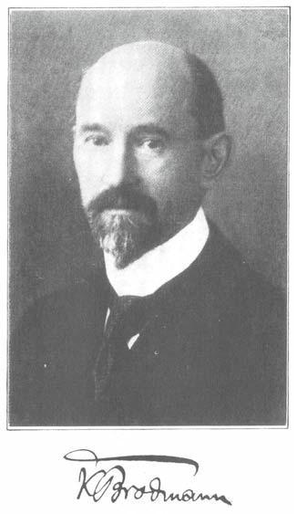 Korbinian Brodmann (1868-1918) was a German neurologist who became famous for his work on the cytoarchitectonic organization of the cerebral cortex. Brodmann's parcellation of the human cortex into about 44 areas (there were some missing numbers) is far from uncontroversial. For example, the Economo and Koskinas atlas published in 1925 distinguishes 107 different areas, and the Vogts thought there were more than 200. Bailey and von Bonin (1951) criticized the proliferation subdivisions, calling it the "crazy pavement" school of cortical research, and recognized less than half the number of areas than Brodmann.
  Despite all this disagreement, and despite the fact that the delineation of areal boundaries in the classical work is highly subjective, it has nonetheless become very common in functional imaging studies to report Brodmann area (BA) numbers associated with activation foci. I have to admit that I caved to this practice. When I first started publishing fMRI studies I didn't bother with BA numbers, or even standardized x, y, z coordinates. I preferred instead to simply show the activations of individual subjects overlaid on their own brain and to describe the location of activations in terms of sulcal and gyral landmarks. Increasingly, reviewers demanded that we report standardized coordinates and BA numbers associated with our activation foci "to allow comparison to other published research." There is certainly some benefit to this practice (it allows for meta-analyses, for example), but still there is a lot of error in the normalization process, and even more in the "alignment" of these localizations to cytoarchitectonic areas defined subjectively 100 years ago. We started reporting coordinates and BA numbers anyway.
I was pleasantly surprised recently when a reviewer criticized a paper I submitted that reported BA numbers. Correctly, the reviewer pointed out that you can't claim that an activation is in a given cytoarchitectonic field without gathering histological data on your subjects. I actually defended the practice in my response saying that the BA numbers were fairly standard in the field, and not interpreted seriously but were simply used as a convenient shorthand. The reviewer argued that we shouldn't follow a bad convention for its own sake and suggested that we remove all reference to BA numbers. S/he was exactly right, and so we gladly accommodated the request! Instead of BA numbers, we simply referred to the activation locations of the group averaged data according to their anatomical position on the normalized brain. (Shhh! Yes, I know there is probably just as much error in group averaging and overlaying on a "standard" brain, but the reviewer was apparently OK with that. Anyway, the point of our paper was not the precise location of the activation but the relation between activations in various conditions.))
Another reviewer comment on a different manuscript criticized our use of an average brain template (the fuzzy-looking MNI average brain) on which we overlaid our activation foci. The image was deemed "poor quality" because you couldn't identify detailed anatomy of the anatomical image. We argued that the fuzzy image probably better reflects the error in the overlay than a high-resolution image that can give the reader a false sense of localization security. We'll see whether the reviewer buys this argument...
All of this has me thinking about the usefulness of group-based, normalized localization practices in functional imaging generally, and the use of Brodmann areas in particular. With respect to the latter, I think it is important to remember that BA number localizations shouldn't be taken literally. As Ted Jones pointed out in a recent book review:
Cortical architecture can only be given functional meaning when correlated with data of a functional character derived using complementary techniques, preferably from the same brain. -Jones (2008)
Without this validation, BA numbers are just shorthand for referring general brain areas. Maybe it's time to give them up. Similar arguments could be made regarding group based localizations. Because of the error in the normalization process itself -- I've seen an individual subject activation focus jump from one sulcus to another after normalizing the subject's brain image -- as well as error associated with cross subject variability, we have to interpret normalized, localizations as very approximate. Single-subject localizations using the subject's own brain image is the only way to get close to valid localization in functional imaging. But even here we have to worry about localization error inherent in the BOLD signal where peak signal can be displaced from the actual site of brain activity, as well as error in the functional-anatomical co-registration.
In general, I think the field is much too localization oriented. We have fooled ourselves into thinking we can localize a group activation to within millimeters. Overlaying group activations on a single high-resolution "standard" brain further promotes the illusion. Perhaps it is this localization illusion that has many of us thinking in terms of the function of area x versus area y. This, of course, is the wrong way to think about brain function. Again, Ted Jones provides an instructive reminder in this new age of localization-based neuroscience:
No cortical area is an isolated entity in which a single function is represented. Nor, contrary to many current views, does it merely form one step in a hierarchy of areas proceeding onwards and upwards to some defined or imagined higher function. While there are definite streams of cortico–cortical connections that proceed in identifiable ways from area to area in the cortex, no area is without feedback connections and no area is without re-entrant connections from the thalamus. -Jones (2008)
References
Bailey P, Von Bonin G. The isocortex of man. Urbana, IL: University of Illinois Press; 1951.
E. G. Jones (2008). Cortical maps and modern phrenology Brain, 131 (8), 2227-2233 DOI: 10.1093/brain/awn158
A. Schleicher, N. Palomero-Gallagher, P. Morosan, S. B. Eickhoff, T. Kowalski, K. de Vos, K. Amunts, K. Zilles (2005). Quantitative architectural analysis: a new approach to cortical mapping Anatomy and Embryology, 210 (5-6), 373-386 DOI: 10.1007/s00429-005-0028-2
Lazaros C. Triarhou (2007). The Economo-Koskinas Atlas Revisited: Cytoarchitectonics and Functional Context Stereotactic and Functional Neurosurgery, 85 (5), 195-203 DOI: 10.1159/000103258
Friday, September 26, 2008
Frontiers in Human Neuroscience
 I just submitted my first paper to the new open-access journal, Frontiers in Human Neuroscience. This journal is part of a constellation of "first-tier" specialty neuroscience "Frontiers in" journals (Frontiers in... Molecular Neuroscience; ...Neuroanatomy; ...Systems Neuroscience; etc.). Prominent articles from these specialty journals are selected for review-style elaboration in the "second-tier" Frontiers in Neuroscience journal. The choice of the term "second-tier" is unfortunate. It implies a less-than-top-notch journal, when in fact the intention is that Frontiers in Neuroscience will be the flagship journal that reaches the broadest audience and has the highest impact.
 I'm usually very hesitant about submitting papers to new untested journals, and in general I think we need new journals just about as bad as we need more greenhouse gases, but I'm on the editorial board of this one and despite my inclusion :-) the board is very strong. So Corianne Rogalsky and I decided to give it a go with her paper on Broca's area, sentence comprehension, and working memory. Unlike most submission/editorial/review processes, I LOVED the experience with Frontiers. We submitted on August 10, and as you can see from my previous post, the paper is already accepted -- and most of that delay time was due to us working on some of the reviewers' suggested analyses of the dataset. So they are fast. The review process itself is interactive, which I find very cool and useful. Reviewers post their comments online and you respond to each issue, also online, in a response field. When you have responded to all comments, you click "done" and this notifies the editor and reviewers that you have responded. They can then reply to your responses if needed, and so on. Russ Poldrack handled the editorial work very efficiently and constructively.
Who knows whether the Frontiers journals will continue their early success, but they are off to a great start. I suppose it is up to us. If we submit to and read a journal regularly, it will flourish. Based on my initial experience, I think this is one journal I'd like to see survive.
Thursday, September 25, 2008
Thalamus? Yes. Basal ganglia? Nope.
A new electrophysiological study just published in Neuron, based on a VERY unusual type of data, provides support for the view articulated by Crosson (1985) and Nadeau & Crosson (1997) (of all the subcortical regions that could matter, it's the thalamus that actually matters for sentence processing) and not such good news for Michael Ullman (declarative/procedural model: of the subcortical regions, the basal ganglia matter the mostest).
A clinical group from Berlin (the Charité Hospital), led by Fabian Klostermann, and a bunch of collaborators (including psycholinguists Angela Friederici, Anja Hahne, and Doug Saddy) studied patients (22 of them) with implanted electrodes for Deep Brain Stimulation (DBS). In DBS, electrodes are implanted to manage, for example, severe tremor, Parkinson's disease, and dystonic diseases. Although these electrodes are placed to deliver electrical stimulation in specific locations, they can also be used for recording.
Electrophysiological data were acquired in these subjects both from the DBS electrodes and scalp electrodes. The DBS placement was in the ventral intermediate nucleus (thalamus); the subthalamic nucleus, and the globus pallidus internus (basal ganglia). Subjects were presented with "the usual", namely syntactic violations eliciting ELAN/LAN and P600 patterns and semantic violations eliciting N400 responses.
All three patient groups showed the standard pattern of ERP responses for the scalp recordings, i.e. ELAN from the left front (F7) and early, N400 and P600 from posterior electrodes, typically Pz. OK, so far so good. What about the subcortical recordings?
The data from VIM show vigorous responses for both syntactic and semantic experimental conditions. However, the recordings from STN and GPi show nothing ... So that's not so great for the model that argued for a central role for the basal ganglia. The authors go pretty directly against Michael Ullman's declarative/procedural model. A large chunk of the discussion is devoted to arguing against Michael's position. I'd be curious to hear his response -- i.e. how do you defend the DP model in light of such findings?
I find the most interesting part of the data the timing between the cortical and thalamic responses. The thalamic responses must be driven by cortical (or other top-down) inputs), because they occur later than the cortical ones ... So what is being fed down to the thalamus? And why?? The authors have some speculations about this, but the story is undeveloped. In any case, since the thalamus receives an enormous number of cortical inputs, the phenomenon merits some attention.
The paper is certainly worth taking a look at because:
-- it's a very unusual source of data
-- the problematic data for the declarative/procedural model merit some thought
-- the timing between the cortical and the thalamic responses point to an interesting relation between these regions. since it can be bottom-up mediated .... what's going on? Attentional gating? Perceptual re-evaluation? Enhancement of those parts of the input that violate local expectations/predictions?
Broca's area, sentence comprehension, and working memory
Broca's area shows a "sentence complexity" effect. It responds more during the comprehension of object relative (OR) constructions than easier to process subject relative (SR) constructions:
OR: The man that the boy pushes is wearing a red shirt
SR: The man that pushes the boy is wearing a red shirt
What is driving the complexity effect? Presumably it is some form of working memory. In the case of OR sentences, you have to hold two items in memory -- the man, the boy -- before you get to the verb that tells you the role these items will play in the sentence. In SR sentences, you only have one item in memory -- the man -- before you encounter the verb which establishes the role of the noun phrase.
But what is the nature of this working memory process? It is a syntactic-specific form of working memory? Or is it an ordinary, domain-general form of working memory, like Baddeley's phonological loop?
David Caplan and colleagues (2000) addressed this issue a while ago in an imaging study that tested for the existence of a sentence complexity effect in Broca's area under conditions of articulatory suppression (subject was continuously articulating a phrase). This manipulation effectively eliminates articulatory rehearsal, a major component of phonological working memory. If the sentence complexity effect in Broca's area was due to phonological working memory -- the articulatory rehearsal component in particular -- then no complexity effect should be evident during articulatory suppression. Caplan et al. reported a complexity effect in Broca's area even during articulatory suppression, however, and so they concluded that the effect was due to "processing syntactic forms themselves" rather than to articulatory rehearsal/phonological working memory.
I like Caplan et al. study, but it doesn't convince me fully that at least part of the complexity effect isn't due to phonological working memory. For one thing, they used written stimuli, and I'm never convinced that reading generalizes to auditory comprehension -- my primary interest. For another thing, the activation focus in Broca's area was very anterior, likely in the pars triangularis/BA45, not in the pars opercularis/BA44 which is more likely a site involved in articulatory rehearsal. Maybe a good chunk of the suppression effect in "Broca's area" -- usually defined as the combination of the two areas noted above -- IS driven by plain old articulatory rehearsal.
Former TB West grad student, Corianne Rogalsky, decided to find out. We first conducted a simple behavioral experiment where undergrads were asked to comprehend sentences either during articulatory suppression, or while performing a control secondary task, finger tapping. During both secondary tasks, subjects were basically at ceiling for active sentences, passive-voice sentences, and SR sentences, but performance dropped off for OR sentences, as expected. Importantly, performance was significantly worse on OR sentences during articulatory suppression compared to finger tapping, indicating that articulatory suppression was interfering with "complex" sentence comprehension above and beyond the general effects of performing two tasks at once. So, articulatory rehearsal seems to be involved in comprehending OR sentences.

What is the role of Broca's area in these effects? We used fMRI to find out. Subjects listened to SR or OR sentences and made semantic plausibility judgments about them during articulatory suppression, finger tapping, or without any secondary task. We also mapped the activation patterns associated with the secondary tasks alone. Without any concurrent task, we found a robust sentence complexity effect in Broca's area, that included both the pars triangularis and pars opercularis:

During articulatory suppression, much of the complexity effect disappeared, leaving only a focus in the pars triangularis similar to Caplan et al.'s finding:

Somewhat unexpectedly, finger tapping also led to an elimination of much of the complexity effect activation in Broca's area, although with a very different distribution. Now the complexity effect is evident in the pars opercularis:

This pars opercularis focus, but not the triangularis focus, was also strongly activated by the articulatory suppression task alone, indicating that this more posterior portion of Broca's region is the IFG correlate of articulatory rehearsal, as previous work has indicated.
So what does all this mean? First, we can conclude that plain old, domain-general articulatory rehearsal contributes to sentence comprehension under high load conditions (i.e., OR sentences), and at least part of the sentence complexity effect in Broca's area is attributable solely to articulatory rehearsal, namely the pars opercularis part. Second, we replicated Caplan's result showing that a portion of Broca's area, the pars triangularis, persists in showing a sentence complexity effect even during articulatory suppression. However, this effect was eliminated during concurrent finger tapping, possibly arguing against a syntactic specific interpretation of the sentence complexity effect in the pars-T. Oddly though, finger tapping alone didn't activate the pars-T, it just reduced the amplitude of the response to the OR sentences (without an obvious drop in behavioral performance). So the story on the pars opercularis seems clear: it supports articulatory rehearsal, which in turn supports the processing of high load sentences. But the story on Mr. T is mysterious. We're open to suggestions.
References
David Caplan, Nathaniel Alpert, Gloria Waters, Anthony Olivieri (2000). Activation of Broca's area by syntactic processing under conditions of concurrent articulation Human Brain Mapping, 9 (2), 65-71 DOI: 10.1002/(SICI)1097-0193(200002)9:23.0.CO;2-4
Rogalsky C, Matchin W and Hickok G (2008) Broca's area, sentence comprehension, and working memory: an fMRI study. Front. Hum. Neurosci. doi:10.3389/neuro.09.014.2008
Tuesday, September 16, 2008
Pulvermuller = Wernicke-Lichtheim
The traditional view has been that language primarily involves two core areas in the left inferior frontal and superior temporal cortex. these were considered modules for language production and comprehension, respectively (Lichtheim,1885; Geschwind, 1970). With the advent of neuroimaging, however, the evidence became overwhelming that, apart from classic core language areas, multiple supplementary areas are also active during language processing, suggesting widely distributed corteial system as the basis of language in the brain (Pulvermuller, 1996...).
-Pulvermuller et al. 2005, p. 794.
Many of Friedemann Pullvermuller's papers start out with a statement, such as that above, which contrasts the old-school "modular" 19th century models of Wernicke and Lichtheim, with "modern" distributed accounts. I've commented on Pulvermuller's misrepresentation of the classic view on semantics in a previous post. Now that I've had to read, several times, how the modern distributed account contrasts so sharply with the classical view, I think it is time to point out that the proposed model of Pulvermuller is identical to that proposed by Wernicke and Lichtheim.
Here is Pulvermuller's Figure 1 from his 1996 paper (Pulvermuller, 1996):

The caption reads,
Sketch of a cortical network that may represent a phonological word form. Simultaneous activity of neurons in the persylvian areas during articulation of word forms may lead to strong coupling of coactivated neurons. Circles represent local neuron clusters, and lines represent axonal connections between such clusters. (p.318)
Notice that the network clusters are centered on classic Broca's and Wernicke's areas. Now, here is Wernicke's Figure 3 (Wernicke, 1874):

Wernicke writes,
...let a be the central termination of the acoustic nerve... Let b stand for the representation of movements in the cerebral cortex which are necessary for the production of sound, connected with a by association fibers ab..."
Save for the fact that Wernicke drew his model (inexplicably) on a right hemisphere, Pulvermuller seems to interpret this diagram quite literally:
According to 19th century neurologists, language is located in two small centers in the language-dominant hemisphere (Broca, 1861; Wernicke, 1874). (Pulvermuller, 1996, p. 317)
However, Wernicke clearly did not intend the diagram to be taken so literally:
The entire region of the first convolution, which circles around the fossa Sylvii serves in conjunction with the insular cortex as a speech center. The first frontal convolution, which is a motor area, is the center of representations of movement; the first temporal convolution, a sensory area, is the center for sound images.
So Wernicke actually proposed a more distributed region of perisylvian cortex as the seat for sensory and motor word images than Pulvermuller proposes for his homologous phonological word forms (if we take Pulvermuller's figure literally).
Pulvermuller claims that the two language centers become "coupled" via simultaneous activity. What does Wernicke claim? Discussing the neural basis of voluntary movement generally, Wernicke (1874) writes,
...the use of these pathways, i.e., the ready transmission of excitation along them, is no accident; rather it can easily be demonstrated that two places in the brain that have once functioned together regularly remain associated.
In a later publication, Wernicke (1900) is crystal clear about his "Hebbian learning" proposal:
During sensory perception two regions within a projection field (or two regions of two differentprojection fields…) become activated simultaneously by the concurrent arrival of external stimuli in the projection fields. Each time this happens, the connection pathway that lies between the two areas begins to resonate, so to speak, with the active regions. The more often this process is repeated, the smaller the resistance to propagation becomes, and the pathway is ground out, or as it is called nowadays, strengthened (p. 34). (Quoted from Gage & Hickok, 2005)
Ok, so for core sensory-motor functions of speech/language, i.e., phonological functions, as it is called nowadays, Wernicke and Pulvermuller are on exactly the same page. What about semantics? Here is Pulvermuller's Figure 3:

The caption for this figure is:
Cell assemblies representing concrete nouns that evoke strong visual associations (visual words) may be distributed over perisylvian and visual cortices. In contrast, cell assemblies representing action verbs that refer to motor activities usually performed by humans (motor words) may include neurons in perisylvian and motor cortices. (p. 320)
So the idea is that the "pre-semantic" phonological aspects of a word are stored in Broca's and Wernicke's area, whereas the concepts that these words refer to -- their visual and motor associations, for example -- are stored in a distributed fashion in other areas according to the type of associations these words have.
What is Wernicke's position on this issue? From his 1874 monograph:
The concept of the word “bell”, for example, is formed by the associated memory images of visual, tactual and auditory perceptions. These memory images represent the essential characteristic features of the object, bell. (Quoted from Gage & Hickok, 2005)
And from his 1885 piece:
… the memory images of a bell … are deposited in the cortex and located according to the sensory organs. These would then include the acoustic imagery aroused by the sound of the bell, visual imagery established by means of form and color, tactile imagery acquired by cutaneous sensation, and finally, motor imagery gained by exploratory movements of the fingers and eyes. (Quoted from Gage & Hickok, 2005).
So Wernicke and Pulvermuller are again on the same page (paragraph even!) when it comes to the regions that house the sensory and motor associations of words. What about Lichtheim? Here is Lichtheim's famous Figure 1 "house model"):

Ignore the "information flow" arrows. Although this depiction is often used to represent the Wernicke-Lichtheim model, Lichtheim actually rejected it in the text of the article. But the main point here is the "concept center" denoted by point B. Did Lichtheim believe concepts were stored in a modular, dedicated concept "center"? Not at all. Lichtheim writes,
Though in the diagram B is represented as a sort of centre for the elaboration of concepts, this has been done for simplicity's sake; with most writers, I do not consider the function to be localised in one spot of the brain, but rather to result from the combined action of the whole sensorial sphere. (p. 477).
Lichtheim's Figure 7 in the same paper illustrates this distributed model.

Notice the distributed nature of the conceptual representation, just like Wernicke and Pulvermuller.
So despite claims to the contrary, Pulvermuller's model of the functional anatomy of language is essentially a recapitulation of the classical model. The only difference seems to be in the number of circles that are drawn in the graphic depictions of the respective models.
References
N GAGE, G HICKOK (2005). Multiregional Cell Assemblies, Temporal Binding and the Representation of Conceptual Knowledge in Cortex: a Modern Theory by a “Classical” Neurologist, Carl Wernicke Cortex, 41 (6), 823-832 DOI: 10.1016/S0010-9452(08)70301-0
FRIEDEMANN PULVERMÜLLER (1996). Hebb's concept of cell assemblies an the psychophysiology of word processing Psychophysiology, 33 (4), 317-333 DOI: 10.1111/j.1469-8986.1996.tb01057.x
Friedemann Pulvermuller, Olaf Hauk, Vadim V. Nikulin, Risto J. Ilmoniemi (2005). Functional links between motor and language systems European Journal of Neuroscience, 21 (3), 793-797 DOI: 10.1111/j.1460-9568.2005.03900.x
Wernicke, C. (1874). The symptom complex of aphasia: A psychological study on an anatomical basis.
Thursday, September 11, 2008
Lipstick on a pig: a neural perspective
 Abstract. Purpose: To investigate the effects of political party affiliation on the interpretation of metaphoric expressions. Approach: Millions of subjects were exposed to a single metaphor, "You can put lipstick on a pig -- It's still a pig" and were asked to indicate the intended referent of the word, "pig." Data collection and analysis: Response data were collected via tedious monitoring of television news channels, particularly CNN, where interpretations of "pig" were offered (repeatedly) by a handful of "representative" subjects. Each responder was classified as belonging either to the Democratic or Republican party, and interpretations were tabulated. Results: Democrats uniformly interpreted "pig" to refer to John McCain's proposed policies, whereas Republicans uniformly interpreted "pig" to refer to Sarah Palin. Conclusion: metaphor interpretation is strongly influenced by political affiliation.
There's only one problem with this study. In a less biased sample of subjects, including myself, a couple of acquaintances, and the audience at Obama's speech which roared with laughter at the comment and (reportly) began chanting "no more pit bull," even some democrats interpreted pig as referencing Sarah Palin. At the very least (admit it), you interpreted the comment as a reference to her pit bull, hockey mom, & lipstick joke.
Here's why: Brains are pretty good at associative learning. Cream and _____, peanut butter and ____, and now, lipstick and _____ ... pit bulls? Sarah Palin? "Lipstick on a pig" was close enough to trigger the association with Sarah Palin and her lipstick joke. We can't help but think of it.
It turns out as well, that there is some commonality in the brain basis for metaphor interpretation and associative learning. Metaphor interpretation compared to the interpretation of literal sentences consistently activates the left inferior frontal gyrus (BA 45/47 for you brain nerds) (Eviatar & Just, 2006; Rapp et al., 2004; Shibata, et al., 2007; Stringaris, et al. 2007). The same area also seems to be activated when subjects listen to phrases that violate semantic associations, such as She spread her bread with socks relative to phrases that are consistent with previous associations, She spread her bread with butter (Willems et al., 2008). This area is often interpreted as supporting the integration of semantic information. Other studies have shown that this inferior frontal gyrus region is involved in semantic memory for previously presented words: it responds differently to a word if that word was presented to the subject previously, even when days intervene between presentation (Meister et al., 2007). Given this patterns of results, it seems likely that this chunk of frontal cortex is involved in our association of Obama's pig and Palin's pit bull.
Does this mean Obama was calling Palin a pig? Not necessarily. A metaphor isn't restricted to one meaning. It could have been a comment on McCain's more-of-the-same policies, and a jab at the lipstick joke. Whatever the intention, the comment certainly opened the door to Republican critics. Obama should have consulted with a language scientist...
References
Z EVIATAR, M JUST (2006). Brain correlates of discourse processing: An fMRI investigation of irony and conventional metaphor comprehension Neuropsychologia, 44 (12), 2348-2359 DOI: 10.1016/j.neuropsychologia.2006.05.007
Ingo G. Meister, Dorothee Buelte, Roland Sparing, Babak Boroojerdi (2007). A repetition suppression effect lasting several days within the semantic network Experimental Brain Research, 183 (3), 371-376 DOI: 10.1007/s00221-007-1051-8
A RAPP, Leube DT, Erb M, Grodd W, Kircher TT. (2004). Neural correlates of metaphor processing Cognitive Brain Research, 20 (3), 395-402 DOI: 10.1016/j.cogbrainres.2004.03.017
M SHIBATA, J ABE, A TERAO, T MIYAMOTO (2007). Neural mechanisms involved in the comprehension of metaphoric and literal sentences: An fMRI study Brain Research, 1166, 92-102 DOI: 10.1016/j.brainres.2007.06.040.
A STRINGARIS, N MEDFORD, V GIAMPIETRO, M BRAMMER, A DAVID (2007). Deriving meaning: Distinct neural mechanisms for metaphoric, literal, and non-meaningful sentences Brain and Language, 100 (2), 150-162 DOI: 10.1016/j.bandl.2005.08.001
Willems RM, Ozyürek A, Hagoort P. (2008). Seeing and hearing meaning: ERP and fMRI evidence of word versus picture integration into a sentence context. J Cogn Neurosci. 20(7):1235-49.
Wednesday, September 10, 2008
Do right motor cortex lesions cause verb processing impairments?
I'm still looking for compelling evidence that damage to the motor system affects verb processing. TMS data was not convincing, nor was ALS data (see previous posts). Now I'm looking at a lesion study by Neininger & Pulvermuller (2003, Word-category specific deficits after lesions in the right hemisphere. Neuropsychologia, 41:53-70), and I have to admit this is a reasonably impressive result -- but not exactly air tight.
Twelve patients with right frontal lobe damage and left hemiparesis were studied, as well as six patients with damage to right inferior temporal-occipital regions. None of the patients were aphasic, consistent with the fact that the lesions were in the right hemisphere. A group of control patients were also tested. Nouns with strong visual associations and verbs with strong action associations were presented visually and subjects were asked to make a lexical decision on each item. A third category of words was included that they called "bimodal nouns" -- nouns with both strong visual and motor associations.
The basic result was that frontal patients made significantly more lexical decision errors on verbs than on both categories of nouns, whereas the temporal-occipital patients made more errors on visual nouns than on verbs, and on visual nouns than bimodal nouns. Controls showed no differences. In reaction time data, there was an overall difference between bimodal nouns and action verbs, but no other effects. In sum, they got a nice double-dissociation in accuracy data, with frontal lesions impairing action verb judgments and temporal-occipital lesions impairing visual noun judgments. No such effect was seen in the RT data, however.
We could quibble about why the effect wasn't seen in RT data when such an effect would be expected (in fact both the frontal and the temp-occ groups were actually numerically slower on the verb than noun stimuli), or with the fact that three of the right frontal patients were ambidextrous, or with the fact that two of the temp-occ patients had sensory-motor defects. But let's take the result as valid with this set of patients.
There are a couple of problems. One is the varied location of the lesions, particularly in the frontal group. Eight of the 12 "frontal" patients had involvement of the temporal lobe, and seven had parietal lobe involvement. All had damage to the basal ganglia. No individual accuracy data was reported. To what extent can we attribute the pattern of errors to "motor cortex" let alone the frontal lobe? We can't. We can conclude that large middle cerebral artery infarcts in the right hemisphere can cause patients to make lexical decision errors on action verbs relative to nouns. This is interesting, but not exactly solid evidence for the view that motor systems support action word processing.
Another problem, which underscores problem number one, is that TMS data from Pulvermuller's group appears to refute the involvement of right motor cortex in action word processing. In the study highlighted in the previous blog entry, TMS to right motor cortex did not lead to any effect on lexical decision responses, unlike the reported effect in the left hemisphere. Apparently, something else is going on in the case of large MCA lesions.
B Neininger (2003). Word-category specific deficits after lesions in the right hemisphere Neuropsychologia, 41 (1), 53-70 DOI: 10.1016/S0028-3932(02)00126-4
Tuesday, September 9, 2008
TMS to motor cortex affects lexical decision to body-part related words - What does this tell us?
 One of the most impressive demonstrations of the functional relevance of motor cortex to action-word processing comes from a TMS study by Friedemann Pulvermuller and colleagues (2005, European Journal of Neuroscience, 21:793-97). These researchers stimulated motor cortex for hand or leg areas while subjects performed a lexical decision task. TMS to hand areas led to faster reaction times to hand-related words (e.g., pick) than leg-related words (e.g., kick), whereas the reverse held for TMS to leg areas. This seems to be a clear case of motor cortex involvement in motor-related language processing.
 Does this mean that the semantics of the word kick is encoded in the motor representation of kicking? Not at all. All it means is that there is an association between the semantics of the word kick and motor programs associated with kicking. It tells us nothing about the nature of the association. Suppose the lexical/conceptual representation of the word kick is stored in neural ensemble A which is distinct from, but connected to, the motor programs for the action kicking which is stored in neural ensemble M in motor cortex. Stimulation of M results in partial activation of A to which it is connected, thus priming the lexical/conceptual representation, which in turn results in faster RTs.
The simplicity and intuitive appeal of embodied accounts of complex processes make it all too easy to accept empirical results as indisputable evidence in support of such accounts when in fact there is much to dispute.
Friedemann Pulvermuller, Olaf Hauk, Vadim V. Nikulin, Risto J. Ilmoniemi (2005). Functional links between motor and language systems European Journal of Neuroscience, 21 (3), 793-797 DOI: 10.1111/j.1460-9568.2005.03900.x
Monday, September 8, 2008
Verb processing deficits in motor neuron disease (ALS/Lou Gehrig's disease)
Embodied cognition views of language processing hold that motor systems are central in processing linguistic forms that involve action-based information. This view predicts that damage to the motor system should produce deficits in processing verbs. There is plenty of evidence that patients with left frontal lobe damage can have deficits in naming actions. But this result does not mean that action knowledge has been disrupted (a typical interpretation of embodied cognition theorists). It could be that verb naming places higher demands on conceptual or grammatical processing, which in turn leads to greater frontal involvement. The tendency for these grammatical class deficits to hold in naming but not in comprehension (e.g., Hillis, et al. 2002. Cognitive Neuropsychology, 19:523-34) is consistent with this possibility because if action knowledge were disrupted, deficits in comprehension should also exist. In addition, the fact that lesions resulting in verb naming deficits extend well beyond the motor system proper, is also consistent with the source of the deficit extending well beyond action knowledge.
T. H. Bak, D. G. O'Donovan, J. H. Xuereb, S. Boniface, J. R. Hodges (2001). Selective impairment of verb processing associated with pathological changes in Brodmann areas 44 and 45 in the motor neurone disease-dementia-aphasia syndrome Brain, 124 (1), 103-120 DOI: 10.1093/brain/124.1.103
In this context, recent studies of amyotrophic lateral sclerosis (also known as Lou Gehrig's disease in the U.S.) would seem to provide just the sort of evidence needed to prove the validity of the embodied cognition view. ALS is a disorder that severely affects motor neurons. If verb processing deficits can be documented in ALS, both in production and comprehension, this would be strong evidence favoring the embodied cognition viewpoint. Claims to this effect exist in the literature, but how solid are they?
One of the first systematic studies of language deficits in ALS, and the first to examine noun versus verb processing is Bak et al. (2001, Brain, 124:103-120). This is an excellent and thorough study that reports on six patients with a dementia/aphasia presentation of ALS. I'm no expert on ALS and I'm just starting to look at this literature, but this immediately raises questions in my mind. Are there cases of ALS with significant motor neuron degeneration but no aphasic symptoms? If so, this would seem to be good evidence against the embodied cognition view no matter what is found in the aphasic variant. Also, how much does the dementia component contribute to language deficits?
With these caveats in mind, let's consider the main findings from this paper. First, clear evidence of language deficits were found in the six ALS patients. Production was severely impaired as was comprehension as measured by the TROG (Test of Reception of Grammar). Of the patients who could perform a naming test, naming of actions was more impaired than naming of objects. A similar pattern was found on the noun versus verb comprehension test, where performance was 86% correct for nouns and 63% correct for verbs (chance = 50%).
Seems like a nice result for the embodied cognition view. This might be fairly compelling if the damage were restricted to motor and/or premotor areas and no higher-order cognitive deficits existed. But a consistent pathological finding was the involvement of BA 44 & 45. Area 44 is typically thought of as being a premotor area, but 45 seems to have much broader functions, being implicated in syntactic processing, response selection, etc. In addition, at least 5 of the 6 patients had prominent psychiatric and/or executive dysfunction, which not only implicate a broader network of brain system, but could also substantially impact test performance. If verb stimuli are more difficult to process than noun stimuli, the dissociation could simply result from a complexity effect rather than a disruption of action knowledge. A group of 20 Alzheimer patients were tested as a control group. They performed much better on the noun and verb comprehension test (>95%), and the noun-verb difference was "not significant" but numerically, AD patients were worse on verbs than nouns with a reported p-value of 0.075. As AD patients should show the reverse dissociation if anything, this may indicate a task difficulty effect.
In sum, variants of ALS can have language deficits that implicate frontal circuits and may affect verb processing to a greater extent than noun processing. The basis of this effect is unclear however, at least from this paper. It will be fun to dig a little deeper. Let me know if you come across a particularly compelling paper in this respect...
T. H. Bak, D. G. O'Donovan, J. H. Xuereb, S. Boniface, J. R. Hodges (2001). Selective impairment of verb processing associated with pathological changes in Brodmann areas 44 and 45 in the motor neurone disease-dementia-aphasia syndrome Brain, 124 (1), 103-120 DOI: 10.1093/brain/124.1.103
Subscribe to:
Comments (Atom)