Nothing found
It looks like nothing was found for this search. Maybe try one of the links below or a new search?
Random videos

BOBB-456
HUNTC-415
SXMA-001
DVRT-046
MKMP-699
SUN-098
VEC-778
STARS-993 ซับไทย
RCTD-602
IPX-690 ซับไทย
JUFD-914 ซับไทย
URE-125
IESP-737
No image
7K HUNTB-242
SSNI-181 ซับไทย
VEC-532
EMBZ-308

PKPR-024
FKRU-013
JUFD-991
OAE-286
PKPD-318
SONE-718 avซับไทย คาเฟ่ซ่อนพิษ พนักงานติดกับดักยา
PKPD-292
ROE-356
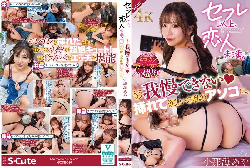
SQDE-026