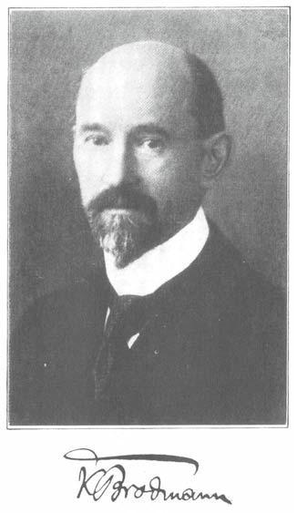
 Korbinian Brodmann (1868-1918) was a German neurologist who became famous for his work on the cytoarchitectonic organization of the cerebral cortex. Brodmann's parcellation of the human cortex into about 44 areas (there were some missing numbers) is far from uncontroversial. For example, the Economo and Koskinas atlas published in 1925 distinguishes 107 different areas, and the Vogts thought there were more than 200. Bailey and von Bonin (1951) criticized the proliferation subdivisions, calling it the "crazy pavement" school of cortical research, and recognized less than half the number of areas than Brodmann.
  Despite all this disagreement, and despite the fact that the delineation of areal boundaries in the classical work is highly subjective, it has nonetheless become very common in functional imaging studies to report Brodmann area (BA) numbers associated with activation foci. I have to admit that I caved to this practice. When I first started publishing fMRI studies I didn't bother with BA numbers, or even standardized x, y, z coordinates. I preferred instead to simply show the activations of individual subjects overlaid on their own brain and to describe the location of activations in terms of sulcal and gyral landmarks. Increasingly, reviewers demanded that we report standardized coordinates and BA numbers associated with our activation foci "to allow comparison to other published research." There is certainly some benefit to this practice (it allows for meta-analyses, for example), but still there is a lot of error in the normalization process, and even more in the "alignment" of these localizations to cytoarchitectonic areas defined subjectively 100 years ago. We started reporting coordinates and BA numbers anyway.
I was pleasantly surprised recently when a reviewer criticized a paper I submitted that reported BA numbers. Correctly, the reviewer pointed out that you can't claim that an activation is in a given cytoarchitectonic field without gathering histological data on your subjects. I actually defended the practice in my response saying that the BA numbers were fairly standard in the field, and not interpreted seriously but were simply used as a convenient shorthand. The reviewer argued that we shouldn't follow a bad convention for its own sake and suggested that we remove all reference to BA numbers. S/he was exactly right, and so we gladly accommodated the request! Instead of BA numbers, we simply referred to the activation locations of the group averaged data according to their anatomical position on the normalized brain. (Shhh! Yes, I know there is probably just as much error in group averaging and overlaying on a "standard" brain, but the reviewer was apparently OK with that. Anyway, the point of our paper was not the precise location of the activation but the relation between activations in various conditions.))
Another reviewer comment on a different manuscript criticized our use of an average brain template (the fuzzy-looking MNI average brain) on which we overlaid our activation foci. The image was deemed "poor quality" because you couldn't identify detailed anatomy of the anatomical image. We argued that the fuzzy image probably better reflects the error in the overlay than a high-resolution image that can give the reader a false sense of localization security. We'll see whether the reviewer buys this argument...
All of this has me thinking about the usefulness of group-based, normalized localization practices in functional imaging generally, and the use of Brodmann areas in particular. With respect to the latter, I think it is important to remember that BA number localizations shouldn't be taken literally. As Ted Jones pointed out in a recent book review:
Cortical architecture can only be given functional meaning when correlated with data of a functional character derived using complementary techniques, preferably from the same brain. -Jones (2008)
Without this validation, BA numbers are just shorthand for referring general brain areas. Maybe it's time to give them up. Similar arguments could be made regarding group based localizations. Because of the error in the normalization process itself -- I've seen an individual subject activation focus jump from one sulcus to another after normalizing the subject's brain image -- as well as error associated with cross subject variability, we have to interpret normalized, localizations as very approximate. Single-subject localizations using the subject's own brain image is the only way to get close to valid localization in functional imaging. But even here we have to worry about localization error inherent in the BOLD signal where peak signal can be displaced from the actual site of brain activity, as well as error in the functional-anatomical co-registration.
In general, I think the field is much too localization oriented. We have fooled ourselves into thinking we can localize a group activation to within millimeters. Overlaying group activations on a single high-resolution "standard" brain further promotes the illusion. Perhaps it is this localization illusion that has many of us thinking in terms of the function of area x versus area y. This, of course, is the wrong way to think about brain function. Again, Ted Jones provides an instructive reminder in this new age of localization-based neuroscience:
No cortical area is an isolated entity in which a single function is represented. Nor, contrary to many current views, does it merely form one step in a hierarchy of areas proceeding onwards and upwards to some defined or imagined higher function. While there are definite streams of cortico–cortical connections that proceed in identifiable ways from area to area in the cortex, no area is without feedback connections and no area is without re-entrant connections from the thalamus. -Jones (2008)
References
Bailey P, Von Bonin G. The isocortex of man. Urbana, IL: University of Illinois Press; 1951.
E. G. Jones (2008). Cortical maps and modern phrenology Brain, 131 (8), 2227-2233 DOI: 10.1093/brain/awn158
A. Schleicher, N. Palomero-Gallagher, P. Morosan, S. B. Eickhoff, T. Kowalski, K. de Vos, K. Amunts, K. Zilles (2005). Quantitative architectural analysis: a new approach to cortical mapping Anatomy and Embryology, 210 (5-6), 373-386 DOI: 10.1007/s00429-005-0028-2
Lazaros C. Triarhou (2007). The Economo-Koskinas Atlas Revisited: Cytoarchitectonics and Functional Context Stereotactic and Functional Neurosurgery, 85 (5), 195-203 DOI: 10.1159/000103258
2 comments:
BA #s are the way to go... The cytoarchitectonic subdivisions of both the thalamus and the neocortex are topographically defined in terms of the variables of phylogenetic age and input specificity. The cortical and thalamic parcellations of Brodmann, von Economo and Hassler are each quantitatively correlated to a specific Cartesian coordinate value designating discrete levels for both age and input basic parameters. The variable of phylogenetic age is represented in the cortex by the five circumferential growth rings demonstrated by Sanides, plus an additional growth ring detected intermediate to the fourth and sixth age levels and designated as “prekoniocortex.” The paleocortex and the archaecortex are the two primordial neocortical precursors that form the mammalian neocortex. In contrast to the arrangement in the planar cortex, six phylogenetically distinct “growth shells” are detected in the three-dimensional thalamus and are designated after the corresponding schematic levels of Rolf Hassler’s paradigm of hexapartition of unit-thalamic inputs. The subthalamus and the epithalamus analogously represent the primordial diencephalic precursors of the mammalian dorsal thalamus, Both the neocortex and the dorsal thalamus evolved in response to the necessity for a more comprehensive blending of inputs from differing neuraxial levels. Unlike the age variable, the parameter of input specificity is most readily apparent in the dorsal thalamus; which is the site of termination for each major forebrain input. Accordingly, the fourteen individual units of the parameter of input specificity are designated after each of the specific input classifications projecting discretely to circumscribed thalamic sectors, An identical complement of input parameter levels also occurs in the cortex by way of thalamic relay across the internal capsule. Furthermore, each thalamic nucleus of specific parameter coordinates directs its main projection to cells of the cortex displaying identical coordinate values, establishing forebrain interconnectivity as an additional function of the dual parameter paradigm. More at www.forebrain.org
This is really hard to follow, even on the forebrain website. Sorry, John, but I don't really get it, either ... Doesn't look like an argument for Brodmann areas, in any case.
Post a Comment