Our mirror neuron course is wrapping up soon. For the final meeting (next monday) we decided that we would all go to the literature and pick our favorite mirror neuron paper and report back to the class on what we learn. I know some students are looking into papers talking about language evolution, others are looking at clinical applications (e.g., autism), so it should be interesting. If any TB readers would like to summarize a favorite paper, please feel free to post a comment!
So what did we learn in this course? One thing I learned is that it is completely unclear what real (i.e., macaque) mirror neurons are doing. There is no evidence whatsoever that mirror neurons in macaque support action understanding. In fact, Rizzolatti has stated that this hypothesis is impossible to test using standard methods (lesion). Might MNs support imitation? Well, macaques are supposed to lack the ability to imitate. If this is true -- and given recent reports the jury may still be out on this -- MNs cannot support imitation. I suggest they simply reflect good old-fashioned sensory-motor associations.
On the human front, we also learned that there is no evidence that the "mirror system" (in quotes because the response characteristics of this system are different from real mirror neurons) supports action understanding, and in the speech domain there is, in fact, good evidence against the view that the "mirror system" is the basis for speech recognition. The "mirror system" may support gesture imitation in humans, which need not involve understanding. But this is just another way of saying that the brain can establish sensory-motor associations, which is certainly not news.
So if there is NO evidence supporting the most flashy claim regarding mirror neurons (action understanding), why has this system gotten so much attention? And why has it been so widely and blindly accepted as truth? I don't know, but I can speculate:
1. The idea has intuitive appeal. Since we all understand our own actions (or at least think we do -- I bet this is debatable), it seems reasonable that we might understand other people's actions by relating them to our own. Be wary of hypotheses with intuitive appeal: they require much less empirical support to convince people that they are true.
2. The idea simplifies a complex issue. Semantics is complex. (See our previous blog discussions.) If we can explain how we "understand" via the response properties of a small population of cells in Broca's area, this avoids all kinds of messy semantic complications.
3. There is a cellular grounding. If the only data regarding the mirror system came from human functional imaging studies, I bet no one would believe it. The fact that a cell in the monkey F5 shows "mirror" properties provides a neurophysiological anchor for human-related speculation, and this goes a long way towards lending credibility.
4. There is a cognitive grounding. The motor theory of speech perception provided an independently motivated theory (even if it is wrong) to back up the general concept of a motor theory of action understanding. It's no accident that the motor theory was referred to in the earliest empirical MN papers.
5. It is easily generalizable. If we understand actions of others by relating them to our own, we might understand speech, emotions, etc., in the same way. Now we have a cellular basis for all sorts of complex systems ranging from speech to empathy, and potential explanations for complex disorders such as autism.
You put all these plusses together and you have a theory that people are willing to impulse-buy without evidence, and without thinking too hard about it.
So the next time you review a paper that states, "This [mirror neuron] research has shown that areas of the brain which subserve motor action production are also involved in action perception and comprehension." (Saygin et al., 2004, Neuropsychologia, 42:1788-1804, p. 1802), note in your review that this claim is speculative and unsupported by empirical evidence. If the authors want to include such a statement, this is fine, but it should be prefaced by "Although there is no evidence to support the theory, mirror neuron research has been used as a basis to speculate that..." Otherwise, the innocent reader will assume that there is an empirical backing to the claim and the speculation will continue to be propagated in the literature as fact. If we take this attitude, the field will be better situated to test the hypothesis rigorously, and if the evidence is supportive, the theory can win over even the skeptics like me. If it is not supported, then we can figure out what this system might be doing. Either way, we make progress.
News and views on the neural organization of language moderated by Greg Hickok and David Poeppel
Wednesday, May 28, 2008
Thursday, May 22, 2008
Comparative neuroscience: Macaque vs. Human brains
We've filled up a fair amount of blog space lately talking about comparative neuroscience -- using functional anatomic data from one species (macaque monkey) to make inferences about the functional organization of neural systems in another (human). I am a big fan of comparative approaches. We can learn a lot from animal models. However, it is important to remember that we are dealing with systems which are not necessarily homologous. This is particularly true when we start talking about higher cognitive functions.
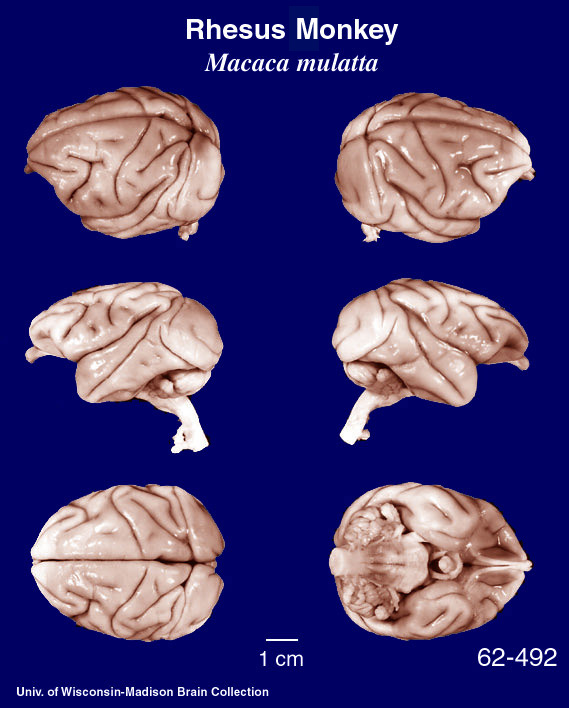
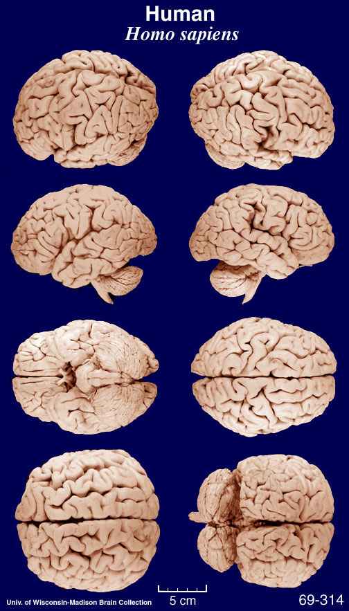
Given that the macaque is a favorite model for much human neuroscience generalization, it is instructive to remind ourselves that the macaque and human brains are rather different on gross examination as the images below make clear. BTW, this is a Macaca mulatta specimen -- a species used in much primate neuroscience research, but not the species in which mirror neurons were discovered which is Macaca nemestrina. But you get the idea.


These images came from the Comparative Mammalian Brain Collections webpage.
Given that the macaque is a favorite model for much human neuroscience generalization, it is instructive to remind ourselves that the macaque and human brains are rather different on gross examination as the images below make clear. BTW, this is a Macaca mulatta specimen -- a species used in much primate neuroscience research, but not the species in which mirror neurons were discovered which is Macaca nemestrina. But you get the idea.


These images came from the Comparative Mammalian Brain Collections webpage.
Wednesday, May 21, 2008
Gesture discrimination in patients with limb apraxia -- evidence for the mirror system?
As hinted at previously, the recent paper by Pazzaglia et al. (2008, J. Neuroscience, 28:3030-41) provides the best evidence I've seen in support of the mirror neuron theory of action understanding. This is a very nice paper, and a brilliant effort to assess the neural basis of gesture discrimination deficits. Unfortunately, there are some complications.
Here's what makes the paper look strong:
Subjects: 41 CVA patients were studied, 33 with left brain damage (LBD) and 8 with right brain damage (RBD). 21 of the LBD subjects were classified as having limb apraxia on assessment.
Stims and task: Subjects viewed video clips of an actor performing transitive or intransitive meaningful gestures. These gestures were performed either correctly or incorrectly. So for example, a correct transitive gesture might show the actor strumming a guitar (with an actual guitar), while the incorrect gesture would show the actor strumming a flute (a semantically related foil), or a broom (an unrelated foil). Subjects made correct/incorrect judgments on each trial.
Results: Lesion analysis revealed some amazing-looking data. This data looks so good, that I thought we'd have to change the blog title to Talking Mirror Neurons! Check it out:

Panel A shows the lesion distribution of patients with limb apraxia (LA+) on the left, and without limb apraxia (LA-) on the right. Notice that the LA+ patients have lesions centered around in the inferior frontal gyrus and inferior parietal lobe.
Panel B shows the subtraction of LA+ vs. LA- which highlights the involvement of posterior frontal areas and inferior parietal areas in LA+ patients. This is consistent with much previous data. So far, so good.
Next they analyzed gesture discrimination performance in a variety of ways.
1. LA+ patients performed significantly worse on gesture discrimination than LA- patients.
2. Significant positive correlations were found between measures of gesture production and gesture discrimination "demonstrating a clear relationship between performing and understanding meaningful gestures" (p. 3034).
3. A cluster analysis was performed on the LA+ patients based on their discrimination performance. This analysis revealed that not all LA+ patients had gesture discrimination deficits, however. In fact seven of the 21 LA+ patients were classified as having no deficit in understanding gestures. So while there is a correlation between production and understanding, these abilities do dissociation as much previous work has shown.
Now on to the really amazing data -- the lesion analysis comparing the LA+ patients with vs. without gesture recognition deficits (+GRD vs. -GRD):

Panel A shows the lesion distribution of these two groups, and Panel B shows the subtraction. It couldn't come out cleaner: patients with limb apraxia and gesture discrimination deficits (LA+[GRD+]) have lesions primarily affecting the inferior posterior frontal gyrus, whereas patients with limp apraxia but without gesture discrimination deficits (LA+[GRD-]) have lesions affecting the posterior parietal lobe.
Further, voxel-based lesion-symptom mapping analysis in all 33 LBD patients revealed that performance on the gesture discrimination task correlated with damage to voxels in the inferior frontal gyrus:

Wow! Mirror neurons rule! Is this solid evidence for the role of the posterior IFG in gesture understanding or what!
Maybe "what." Here's two reasons to be suspicious of the findings.
1. The task effectively used a signal detection type paradigm: view a stimulus and decide whether it is signal (correct gesture) or noise (incorrect gesture). The proper way to analyze such data is to calculate d-prime, as this provides an unbiased measure of discriminability. Unfortunately, most of the behavioral analyses, and all of the lesion analyses used uncorrected error rates: a score of 1 was given for hits and for correct rejections, and zeros were given for false alarms and misses. This scoring produces biases, particularly when there are more "noise" trials than "signal" trials, as there were in this study which used a 2:1 noise:signal ratio.
Here's a demonstration using an extreme scenario. Assume three subjects cannot discriminate signal from noise at all but have different response biases:
Subject #1: neutral response bias
10 correct trials: neutral response bias = 5/10 correct
20 incorrect trials: no response bias = 10/20 correct
Overall score = 15/30 correct = 50% accuracy (a valid result)
Subject #2: 100% "yes" response bias
10 correct trials: 100% "yes" response bias = 10/10 correct
20 incorrect trials: 100% "yes" response bias = 0/20 correct
Overall score = 10/30 correct = 33% correct
Subject #3: 100% "no" response bias
10 correct trials: 100% "no" response bias = 0/10 correct
20 incorrect trials: 100% "no" response bias = 20/20 correct
Overall score = 20/30 correct = 67% correct
So you get different accuracy scores depending on the subject's response bias, independently of his/her actual ability to discriminate signal from noise. Of course these biases may not be THIS extreme in practice, but they will find their way into the data, and taint the results -- this is the reason for the d-prime statistic. Is it possible that the frontal and parietal regions are correlating with different response biases? Some people have argued that frontal cortex is important for response selection...
Upshot: the lesion analyses reported in this paper absolutely have to be redone using d-prime measures instead of overall accuracy. Until then, we cannot be sure how much of these findings are driven by response bias.
Some of you are still very confident that even with d-prime, the results will look similar. I wonder myself whether it will change much. But there's another reason to be suspicious...
2. Figure 5. This figure shows the correlation between the percentage of lesioned tissue in the IFG and overall score on gesture recognition, broken down by gesture type, transitive and intransitive. GRD+ and GRD- patients are included in these correlations. Both correlations are reported as significant, with r-values > . 55; not bad. But it is clear from looking at the graphs that the effect is driven by the -GRD group, who all cluster together.

But isn't that part of point? Patients without gesture recognition deficits don't have much damage to IFG, so they should cluster together, right? True, but it should also be the case that for those patients WITH gesture recognition deficits there should still be a strong correlation with amount of lesioned tissue in IFG (assuming there's enough variance in the lesioned tissue, which there is). However, if you remove the -GRD patients (the square symbols), it looks as though the regression line would be completely flat! This is true even thought the percentage of damaged tissue in the +GRD group ranges from ~5% to ~55%. In fact, eyeballing the top graph you can see that the three patients with the LEAST amount of damage to IFG (~to the amount damaged in the -GRD group!) are performing the same as the four patients who have the MOST IFG damage! Further, all the patients in between these two extremes are distributed in a nebulous cloud that kind of looks like an "X" that is equally wide as it is tall (can you find the cross-over double-dissociation in the X?) If IFG was the critical substrate for gesture recognition, one would expect some kind of pattern here, yet there is none.
It seems quite clear that IFG damage is NOT predicting gesture discrimination deficits. Something else is driving the effect that shows up in the beautiful lesion analyses.
Overall conclusion: Pazzaglia et al. made a valiant effort, and should be commended for looking at their data from so many angles, and for reporting their findings openly and straightforwardly. As it stands, however, the data do not support the mirror neuron theory of action understanding. In fact, the data seem to indicate (i) action production and action understanding dissociate, and (ii) damage to the presumed human homologue of monkey area F5 does not correlate with action understanding deficits.
I would love to see the follow up paper that re-analyzes all the data using d-primes. Maybe then we'll have at least one piece of evidence supporting the mirror neuron theory of action understanding. To date, there isn't any evidence in support of the theory.
Here's what makes the paper look strong:
Subjects: 41 CVA patients were studied, 33 with left brain damage (LBD) and 8 with right brain damage (RBD). 21 of the LBD subjects were classified as having limb apraxia on assessment.
Stims and task: Subjects viewed video clips of an actor performing transitive or intransitive meaningful gestures. These gestures were performed either correctly or incorrectly. So for example, a correct transitive gesture might show the actor strumming a guitar (with an actual guitar), while the incorrect gesture would show the actor strumming a flute (a semantically related foil), or a broom (an unrelated foil). Subjects made correct/incorrect judgments on each trial.
Results: Lesion analysis revealed some amazing-looking data. This data looks so good, that I thought we'd have to change the blog title to Talking Mirror Neurons! Check it out:

Panel A shows the lesion distribution of patients with limb apraxia (LA+) on the left, and without limb apraxia (LA-) on the right. Notice that the LA+ patients have lesions centered around in the inferior frontal gyrus and inferior parietal lobe.
Panel B shows the subtraction of LA+ vs. LA- which highlights the involvement of posterior frontal areas and inferior parietal areas in LA+ patients. This is consistent with much previous data. So far, so good.
Next they analyzed gesture discrimination performance in a variety of ways.
1. LA+ patients performed significantly worse on gesture discrimination than LA- patients.
2. Significant positive correlations were found between measures of gesture production and gesture discrimination "demonstrating a clear relationship between performing and understanding meaningful gestures" (p. 3034).
3. A cluster analysis was performed on the LA+ patients based on their discrimination performance. This analysis revealed that not all LA+ patients had gesture discrimination deficits, however. In fact seven of the 21 LA+ patients were classified as having no deficit in understanding gestures. So while there is a correlation between production and understanding, these abilities do dissociation as much previous work has shown.
Now on to the really amazing data -- the lesion analysis comparing the LA+ patients with vs. without gesture recognition deficits (+GRD vs. -GRD):

Panel A shows the lesion distribution of these two groups, and Panel B shows the subtraction. It couldn't come out cleaner: patients with limb apraxia and gesture discrimination deficits (LA+[GRD+]) have lesions primarily affecting the inferior posterior frontal gyrus, whereas patients with limp apraxia but without gesture discrimination deficits (LA+[GRD-]) have lesions affecting the posterior parietal lobe.
Further, voxel-based lesion-symptom mapping analysis in all 33 LBD patients revealed that performance on the gesture discrimination task correlated with damage to voxels in the inferior frontal gyrus:

Wow! Mirror neurons rule! Is this solid evidence for the role of the posterior IFG in gesture understanding or what!
Maybe "what." Here's two reasons to be suspicious of the findings.
1. The task effectively used a signal detection type paradigm: view a stimulus and decide whether it is signal (correct gesture) or noise (incorrect gesture). The proper way to analyze such data is to calculate d-prime, as this provides an unbiased measure of discriminability. Unfortunately, most of the behavioral analyses, and all of the lesion analyses used uncorrected error rates: a score of 1 was given for hits and for correct rejections, and zeros were given for false alarms and misses. This scoring produces biases, particularly when there are more "noise" trials than "signal" trials, as there were in this study which used a 2:1 noise:signal ratio.
Here's a demonstration using an extreme scenario. Assume three subjects cannot discriminate signal from noise at all but have different response biases:
Subject #1: neutral response bias
10 correct trials: neutral response bias = 5/10 correct
20 incorrect trials: no response bias = 10/20 correct
Overall score = 15/30 correct = 50% accuracy (a valid result)
Subject #2: 100% "yes" response bias
10 correct trials: 100% "yes" response bias = 10/10 correct
20 incorrect trials: 100% "yes" response bias = 0/20 correct
Overall score = 10/30 correct = 33% correct
Subject #3: 100% "no" response bias
10 correct trials: 100% "no" response bias = 0/10 correct
20 incorrect trials: 100% "no" response bias = 20/20 correct
Overall score = 20/30 correct = 67% correct
So you get different accuracy scores depending on the subject's response bias, independently of his/her actual ability to discriminate signal from noise. Of course these biases may not be THIS extreme in practice, but they will find their way into the data, and taint the results -- this is the reason for the d-prime statistic. Is it possible that the frontal and parietal regions are correlating with different response biases? Some people have argued that frontal cortex is important for response selection...
Upshot: the lesion analyses reported in this paper absolutely have to be redone using d-prime measures instead of overall accuracy. Until then, we cannot be sure how much of these findings are driven by response bias.
Some of you are still very confident that even with d-prime, the results will look similar. I wonder myself whether it will change much. But there's another reason to be suspicious...
2. Figure 5. This figure shows the correlation between the percentage of lesioned tissue in the IFG and overall score on gesture recognition, broken down by gesture type, transitive and intransitive. GRD+ and GRD- patients are included in these correlations. Both correlations are reported as significant, with r-values > . 55; not bad. But it is clear from looking at the graphs that the effect is driven by the -GRD group, who all cluster together.

But isn't that part of point? Patients without gesture recognition deficits don't have much damage to IFG, so they should cluster together, right? True, but it should also be the case that for those patients WITH gesture recognition deficits there should still be a strong correlation with amount of lesioned tissue in IFG (assuming there's enough variance in the lesioned tissue, which there is). However, if you remove the -GRD patients (the square symbols), it looks as though the regression line would be completely flat! This is true even thought the percentage of damaged tissue in the +GRD group ranges from ~5% to ~55%. In fact, eyeballing the top graph you can see that the three patients with the LEAST amount of damage to IFG (~to the amount damaged in the -GRD group!) are performing the same as the four patients who have the MOST IFG damage! Further, all the patients in between these two extremes are distributed in a nebulous cloud that kind of looks like an "X" that is equally wide as it is tall (can you find the cross-over double-dissociation in the X?) If IFG was the critical substrate for gesture recognition, one would expect some kind of pattern here, yet there is none.
It seems quite clear that IFG damage is NOT predicting gesture discrimination deficits. Something else is driving the effect that shows up in the beautiful lesion analyses.
Overall conclusion: Pazzaglia et al. made a valiant effort, and should be commended for looking at their data from so many angles, and for reporting their findings openly and straightforwardly. As it stands, however, the data do not support the mirror neuron theory of action understanding. In fact, the data seem to indicate (i) action production and action understanding dissociate, and (ii) damage to the presumed human homologue of monkey area F5 does not correlate with action understanding deficits.
I would love to see the follow up paper that re-analyzes all the data using d-primes. Maybe then we'll have at least one piece of evidence supporting the mirror neuron theory of action understanding. To date, there isn't any evidence in support of the theory.
Monday, May 19, 2008
Apraxia, Gesture Recognition, and Mirror Neurons
This week in our Mirror Neuron course we looked over some papers in the apraxia literature, which seemed to hold the promise of providing evidence in support of the MN theory of action understanding. Specifically, based on some of the abstracts, I thought we would find (i) that disorders to gesture production would be strongly associated with disorders of gesture understanding, and (ii) that damage to the frontal (putative) mirror neuron system should be associated with these production/comprehension disorders.
Instead, and despite the conclusions of some of the authors, the papers we read made a pretty decent case against the MN theory of action understanding.
First of all, it is very clear that deficits in gesture production/imitation dissociate from the ability to comprehend gestures. For example, Table 4 of Tessari et al. 2007 (Brain, 130:1111-26) shows data that includes eight (8) patients (out of a sample of 32 left hemisphere patients), who are performing at 60% correct or less on action imitation, while recognizing actions at 90% or better. Case 27, is a dramatic example, with only 10% correct imitations of meaningful actions and 100% correct on action recognition. It's possible, though, that imitation deficits in such cases result from some relatively peripheral motor control mechanism that is outside the mirror system proper. This is reasonable, but then I have to wonder whether data from say, motor evoked potentials, which are touted as strong evidence for the mirror system (and which might be considered rather peripheral!), should now be discounted for being peripheral to the mirror system. In other words, you can't point to a very peripheral response like MEPs and call it evidence for mirror neurons, and then turn around and explain imitation deficits caused by (non-M1) cortical damage as arising from systems that are peripheral to the mirror system.
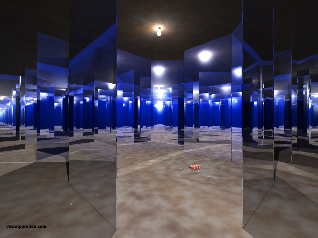
In other, other words, it's time to be explicit about what components of the motor system are part of the mirror system. As it stands, "mirror neuron theory" is effectively untestable because any result just gets folded into the mythology: humans show "mirror" responses for pantomimed gestures (unlike macaque F5 cells)? That's because humans are more sophisticated than monkeys. The STS responds just as selectively (more so!) to perceived actions than F5? That's because it inherits its mirror properties from the real mirror system. Passive viewing of non-action stimuli (rectangles with dots in them) activates the mirror system? That's because of motor imagery associated with the non-action stimuli. Mirror neuron theorizing is starting to remind me of one of those house-of-mirrors attractions at the county fair: Mirrors everywhere, but mostly reflecting nothing.
But I digress... Let's focus on the two papers that seem to make the best case for action understanding deficits associated with lesions to the inferior frontal gyrus.
Saygin et al. studied the ability of aphasic patients to understand action-related pictures. Ok, not exactly action understanding (pictures are static), but let's assume that these pictures induced action percepts/concepts. Subjects viewed actions with the object of the action removed, such as a boy licking an ice cream cone, but with no ice cream cone in his hand. Subjects then had to pick the matching object out of an array of two pictured objects which included the target (ice cream cone) and a distractor. Distractors included (on different trials) semantically related items (cake), "affordance" related items (a bouquet of flowers which is held in a similar manner but not normally licked), or an unrelated item (rooster). A matched verbal version of the task was also administered in which subjects read sentence fragments ("He is licking the _____") and had to pick the correct picture, as above.
The results were quite interesting: (i) performance on the two task was completely uncorrelated (after the one outlier patient was removed), showing that action understanding is domain-specific. (ii) lesion analysis revealed that deficits on the pictorial task were correlated with lesions in the inferior frontal gyrus, whereas deficits on the linguistic task were correlated with a different distribution of lesions that was more posterior and involved the anterior temporal lobe (interesting for other reasons). So both behavioral and brain data show these two tasks dissociate, and pictorial action understanding is associated with the frontal "mirror system." The results were interpreted as support for the mirror neuron theory of action understanding.
But this is an odd theoretical position. Apparently, the ability to understand actions generally, is dependent on the sensory signals that access that action-concept information. That is, having a deficit in the ability to understand actions from action depictions, does not prevent you from accessing action-concepts via a verbal route. Put another way, damage to your mirror neuron system (the frontal lesions associated with pictorial action understanding deficits) leaves action-concepts (action understanding) intact, as evidenced by the fact that you can access them via another input route. Therefore, the representation of action meaning (the understanding part) is not in the motor representation, but is somewhere else. At most the motor system may facilitate access to the meaning of actions. But these data show that the semantics of action is NOT inherent to the motor system. Alternatively, the correlation with frontal lobe structures for this action understanding task, may have more to do with making complex inferences from static ambiguous pictures (have a look at the sample stimulus item in the paper -- the "licking boy" could just as easily be smiling real big, or have a fat lip).
Enough for now. I'll comment in the next post on the strongest evidence we've seen so far for the MN theory of action understanding. (Turns out its not so strong.)
Instead, and despite the conclusions of some of the authors, the papers we read made a pretty decent case against the MN theory of action understanding.
First of all, it is very clear that deficits in gesture production/imitation dissociate from the ability to comprehend gestures. For example, Table 4 of Tessari et al. 2007 (Brain, 130:1111-26) shows data that includes eight (8) patients (out of a sample of 32 left hemisphere patients), who are performing at 60% correct or less on action imitation, while recognizing actions at 90% or better. Case 27, is a dramatic example, with only 10% correct imitations of meaningful actions and 100% correct on action recognition. It's possible, though, that imitation deficits in such cases result from some relatively peripheral motor control mechanism that is outside the mirror system proper. This is reasonable, but then I have to wonder whether data from say, motor evoked potentials, which are touted as strong evidence for the mirror system (and which might be considered rather peripheral!), should now be discounted for being peripheral to the mirror system. In other words, you can't point to a very peripheral response like MEPs and call it evidence for mirror neurons, and then turn around and explain imitation deficits caused by (non-M1) cortical damage as arising from systems that are peripheral to the mirror system.
In other, other words, it's time to be explicit about what components of the motor system are part of the mirror system. As it stands, "mirror neuron theory" is effectively untestable because any result just gets folded into the mythology: humans show "mirror" responses for pantomimed gestures (unlike macaque F5 cells)? That's because humans are more sophisticated than monkeys.
 The STS responds just as selectively (more so!) to perceived actions than F5? That's because it inherits its mirror properties from the real mirror system. Passive viewing of non-action stimuli (rectangles with dots in them) activates the mirror system? That's because of motor imagery associated with the non-action stimuli. Mirror neuron theorizing is starting to remind me of one of those house-of-mirrors attractions at the county fair: Mirrors everywhere, but mostly reflecting nothing.
But I digress... Let's focus on the two papers that seem to make the best case for action understanding deficits associated with lesions to the inferior frontal gyrus.
Saygin et al. studied the ability of aphasic patients to understand action-related pictures. Ok, not exactly action understanding (pictures are static), but let's assume that these pictures induced action percepts/concepts. Subjects viewed actions with the object of the action removed, such as a boy licking an ice cream cone, but with no ice cream cone in his hand. Subjects then had to pick the matching object out of an array of two pictured objects which included the target (ice cream cone) and a distractor. Distractors included (on different trials) semantically related items (cake), "affordance" related items (a bouquet of flowers which is held in a similar manner but not normally licked), or an unrelated item (rooster). A matched verbal version of the task was also administered in which subjects read sentence fragments ("He is licking the _____") and had to pick the correct picture, as above.
The results were quite interesting: (i) performance on the two task was completely uncorrelated (after the one outlier patient was removed), showing that action understanding is domain-specific. (ii) lesion analysis revealed that deficits on the pictorial task were correlated with lesions in the inferior frontal gyrus, whereas deficits on the linguistic task were correlated with a different distribution of lesions that was more posterior and involved the anterior temporal lobe (interesting for other reasons). So both behavioral and brain data show these two tasks dissociate, and pictorial action understanding is associated with the frontal "mirror system." The results were interpreted as support for the mirror neuron theory of action understanding.
But this is an odd theoretical position. Apparently, the ability to understand actions generally, is dependent on the sensory signals that access that action-concept information. That is, having a deficit in the ability to understand actions from action depictions, does not prevent you from accessing action-concepts via a verbal route. Put another way, damage to your mirror neuron system (the frontal lesions associated with pictorial action understanding deficits) leaves action-concepts (action understanding) intact, as evidenced by the fact that you can access them via another input route. Therefore, the representation of action meaning (the understanding part) is not in the motor representation, but is somewhere else. At most the motor system may facilitate access to the meaning of actions. But these data show that the semantics of action is NOT inherent to the motor system. Alternatively, the correlation with frontal lobe structures for this action understanding task, may have more to do with making complex inferences from static ambiguous pictures (have a look at the sample stimulus item in the paper -- the "licking boy" could just as easily be smiling real big, or have a fat lip).
Enough for now. I'll comment in the next post on the strongest evidence we've seen so far for the MN theory of action understanding. (Turns out its not so strong.)
Friday, May 16, 2008
Happy Birthday to Talking Brains
Speaking of birthdays, here's a picture of me with a pretty creative birthday cake given to me by my lab a few years ago.

Wednesday, May 14, 2008
Spoken Word Memory Traces within the Human Auditory Cortex
This looks like an interesting study using fMRI repetition suppression methods to identify neural networks involved in spoken word recognition. It appears such effects were found bilaterally in the superior temporal gyrus. Worth a close look!
Spoken Word Memory Traces within the Human Auditory Cortex Revealed by Repetition Priming and Functional Magnetic Resonance Imaging
Pierre Gagnepain, Gael Chetelat, Brigitte Landeau, Jacques Dayan, Francis Eustache, and Karine Lebreton
J. Neurosci. 2008;28 5281-5289
Spoken Word Memory Traces within the Human Auditory Cortex Revealed by Repetition Priming and Functional Magnetic Resonance Imaging
Pierre Gagnepain, Gael Chetelat, Brigitte Landeau, Jacques Dayan, Francis Eustache, and Karine Lebreton
J. Neurosci. 2008;28 5281-5289
Tuesday, May 13, 2008
Mirror Neuron Course: Reading set #5 - Apraxia and gesture comprehension
So far we have not found any evidence to support the claim that the mirror neuron system is the "basis" of action understanding. In fact, all of the evidence discussed so far, refutes this claim. Let's see if we can find some evidence in the apraxia literature. Apraxic patients should have a deficit in comprehending gestures, and the underlying lesions should implicate the mirror system. Scanning some of the abstracts of the reading list below suggests the evidence is mixed.
Pazzaglia M, Smania N, Corato E, Aglioti SM.
Neural underpinnings of gesture discrimination in patients with limb apraxia.
J Neurosci. 2008 Mar 19;28(12):3030-41.
PMID: 18354006 [PubMed - indexed for MEDLINE]
Halsband U, Schmitt J, Weyers M, Binkofski F, Grützner G, Freund HJ.
Recognition and imitation of pantomimed motor acts after unilateral parietal and premotor lesions: a perspective on apraxia.
Neuropsychologia. 2001;39(2):200-16.
PMID: 11163376 [PubMed - indexed for MEDLINE]
Buxbaum LJ, Kyle K, Grossman M, Coslett HB.
Left inferior parietal representations for skilled hand-object interactions: evidence from stroke and corticobasal degeneration.
Cortex. 2007 Apr;43(3):411-23.
PMID: 17533764 [PubMed - indexed for MEDLINE]
Saygin AP, Wilson SM, Dronkers NF, Bates E.
Action comprehension in aphasia: linguistic and non-linguistic deficits and their lesion correlates.
Neuropsychologia. 2004;42(13):1788-804.
PMID: 15351628 [PubMed - indexed for MEDLINE]
Tessari A, Canessa N, Ukmar M, Rumiati RI.
Neuropsychological evidence for a strategic control of multiple routes in imitation.
Brain. 2007 Apr;130(Pt 4):1111-26. Epub 2007 Feb 9.
PMID: 17293356 [PubMed - indexed for MEDLINE]
Subscribe to:
Posts (Atom)